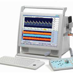

CURRY 8 is an advanced software tool offering large amounts of functionality. It is described in detail in the CURRY 8 User Guide and the CURRY 8 Installation and Tutorials. In addition to the written documentation, this course is available. CURRY integrates multiple complementary imaging modalities such as EEG, ECoG, MEG, MRI, fMRI, CT. By combining the latest techniques for determining electrical activity in the brain with anatomical and functional imaging, CURRY provides a powerful state-of-the-art method for accurately localizing the source of such activity. CURRY uses the full physical anatomy from MR and CT to provide three-dimensional models of the head and brain, pinpointing the site of activity. CURRY integrates fMRI functional imaging with EEG and MEG source analysis. CURRY can be used on PCs and laptops running Windows 7 or later. It is aimed at functional mapping and diagnosis of brain activity. However, the functionality offered and the evolutionary nature of CURRY also make it suitable for wider application.


- HIGHLIGHTS
- Modular design: buy the features you really need, upgrade easily.
- Integration of functional data (EEG, MEG, ECoG, sEEG, ECG, MCG)
with image data (MRI, fMRI, CT, DTI FA, PET, SPECT). - EEG data acquisition module with online processing capabilities.
- Complete data processing from filtering to source analysis.
- Event support, threshold and template-based event detection.
- Selective averaging (event type or SNR based).
- Artifact detection and reduction (subtraction and projection methods).
- Principal and Independent Component Analysis (PCA, ICA) and filtering.
- Time and frequency domain evaluations.
- Current Source Density (Laplacian).
- Individual realistic head models using Boundary and Finite Element Methods (BEM/FEM).
- Pre-computed realistic BEM head models.
- Dipole fits. Dipole confidence ellipsoids are computed.
- Dipole scans, extended source (patch) scans, MUSIC and beamformer scans.
- Current density analysis, extended sources, Lp norms, sLORETA, SWARM.
- Sensor and source coherence, group statistics.
- Export of results in Excel, MATLAB, and SPM formats.
- Interface for data processing in MATLAB.
- Automation and batch processing.
- Report generator.
- Multi-document, workflow-enabled Windows user interface.
- Multi-core support using thread-based multitasking and parallelization.
- Hardware-accelerated real time rendering of 3D scenarios.
- Context-sensitive help system.
CURRY reads a broad variety of EEG, MEG, digitizer, and image file formats. CURRY is backward compatible: It will read all legacy CURRY input files, as well as all CURRY 3 and later output files. Supported EEG data formats include Neuroscan, Compumedics, BioSemi, BESA, BrainVision, EBS, EDF, EGI, Micromed, Bio-Logic, Stellate, XLTEK, Nervus, Nexstim, Nicolet, Nihon-Kohden, Persyst, Telefactor, raw binary and raw ASCII. Supported MEG data formats include Elekta Neuromag, Yokogawa, 4D-Neuroimaging (BTI), CTF,
Philips, BESA, raw binary and raw ASCII. Supported image data formats include DICOM, nifti, SPM, Analyze, Freesurfer, BrainVoyager, Siemens, GE, ACR-NEMA, JPEG, PNG, BMP, GIF, TIFF, raw binary and raw ASCII.


“Complete, Integrated and Simple system to record high quality of EEG and ERP Simultaneously with fMRI”
MicroMagLink system; the next generation technology for simultaneous EEG/fMRI data acquisition – simple The MicroMagLink is based on the concept that often the simplest solution is best. The MicroMagLink keeps all active electronics outside the MRI chamber; all that is placed in MRI chamber is a special electrode cap, a cable, and a fiber optic pulseometer to provide timing for ballistocardiogram suppression. By keeping the active electronic components in the control room, we provide the best possible signal amplification without any compromises.
The MicroMagLink system consists of a precisely engineering electrode placement system with cabling that includes inline RF filtering to ensure that all signals recorded inside the MRI are uncontaminated by any sources outside the shielded MRI chamber. Using our SynAmps RT amplifier, data sampling rates up to 20,000 kHz perchannel are possible. But with the inherent capability of SynAmps amplifiers to synchronize internal clocking to external sources, data sample rates as low as 500 Hzmay be sufficient to provide high quality suppression of gradient sequence artifact.
Following ASTM standards, MicroMagLink electrodes have been engineered and tested to comply with required standards for heating, displacement and torsion up to 3 Tesla. With a single attachment to the penetration panel, the MicroMaglink may routinely be installed and removed quickly. This portability allows a single MicroMaglink to be easily deployed to any magnet in your MRI facility, or used to collect EEG simultaneously with MEG.


Application
- EEG/Evoked Potentials : is compatible with most stimulus presentation system providing a TTL or synchronization pulse output, enabling acquisition of ERP and fMRI simultaneously.
- Sleep : Record your sleep EEG in the MRI and then review with Compumedics Profusion sleep Software
- Epilepsy : EEG recordings can be used to both mark the time of onset of seizures during fMRI recording as well as provide independent evaluation of the site of origin of activities
- Simultaneous EEG/MEG : without compromising the quality of the MEG signal, appropriate RF-Filtering and installation of the core component of the MicroMaglink prevent this extraneous electrical interference from compromising the MEG recordings


Curry NeuroImaging Suite
The MicroMagLink is integrated with the Curry NeuroImaging Suite, which now includes a data acquisition module for capturing EEG data recorded in the MRI. By incorporating data acquisition into the Curry platform, all stages of EEG/ERP data recording and analysis, including source reconstruction, and co-registration with MRI, fMRI and other neuroimaging modalities are handled in a single platform. An advanced set of signal processing methods can be employed both online and offline to suppress both gradient and ballistocardiogram artifact.


The SynAmps RT is seamlessly compatible with all of our other systems. SynAmps RT was designed with specific engineering considerations to provide the best possible recordings in the MRI. SynAmps RT uses the same trigger connector that the original SynAmps and NuAmps use, so there is no need to modify your existing stimulus protocols.
Highlights
-SynAmps RTis a 70 channel amplifier system, consisting of 64 monopolar, 4 bipolar, and 2 high-level input channels (for receiving voltage outputs from other equipment) per headbox. Each channel has a dedicated 24 bit A-to-D converter, to ensure the most accurate sampling available.
-Up to four head boxes can be combined with a single System Unit, creating a 256 channel system. Combine two System Units to create a super-high density system.
-SynAmps RTis a true DC amplifier, allowing you to record slow potentials with no high pass filtering.
-Sampling rates up to 20,000Hz (synchronized across all channels) let you record the fastest activity such as ABRs (maximum low pass filter is 3500Hz).
-Low noise 24 bit A-to-D converters allow for low gain to be used while still maintaining the resolution and a broad dynamic range (making saturation almost impossible).
-Clock synchronizationwith MR scanner triggers allows precise reduction of gradient artifact, without the need for fast sampling rates.
-Active Noise Cancellationensures accurate recordings even in the most hostile electromagnetic environments.
-De-blocking Circuitryallows for recording of TMS and other high intensity stimuli without affecting the integrity of the data.
-Input impedanceof 10 GOhm.
-Common Mode Rejection Ratioof 110 dB.
MagLink system is used for obtaining integrated EEG and ERP recordings inside the fMRI. This second-generation system improves on the original Opti-Link system by increasing the bandwidth and eliminating the pre-amplifier and optical coupling. The passive MagLink system provides the ability to record during the pulse sequence without compromising the raw EEG data. MagLink, in conjunction with Neuroscan amplifiers has successfully recorded EEG data through the pulse sequence in up to 3 Tesla MRI scanners. The CURRY 7 Acquisition and Signal Processing modules provide powerful features for on-line and off-line reduction of the ballistocardiogram and pulse sequence artifact, while retaining the integrity of the EEG. Post-processing tools allow ERP’s to be extracted, which can then be subjected to source localization and compared with the fMRI.
The gradient artifact that is injected into the EEG data can be reduced by a number of different strategies that are included in the CURRY 7 software. Having a variety of data processing methods available is critical to account for the different artifacts that are encountered. The individual artifact signatures vary based on the type of pulse sequence, magnet type and strength, the electrode impedance, even the quality of the shim on a particular day. Neuroscan’s correction methods can correct the EEG data under a variety of parameters.
The SIGNAL PROCESSING license includes SCAN’s signal processing transforms as well as many more advanced signal processing tools based on CURRY’s more complex data processing module. Virtually, signal processing is performed “in place”, without the necessity of creating intermediary files. Functions of this module include all of the online features mentioned above including:
- user-determined re-referencing
- filtering
- baseline correction
- artifact reduction
- averaging
- noise estimation
- PCA and ICA analysis
- spectral analysis including wavelets
- topographic mapping without or without Laplacian derivations,
- sensor/electrode coherence
- and much more.


Stim2 is the next evolutionary step in stimulus presentation and experimental design. Running within the Windows XP (only) environment, Stim2 retains the functionality and timing of the original DOS-based STIM, but with a contemporary user interface and numerous expanded features.
Stim2 is a complete software environment for custom stimulus and task design as well as presentation. The Stim2 system can be integrated in fMRI, MEG, and other functional neuro-imaging applications. The software provides a familiar menu-driven interface in which to design and present visual and auditory stimulation paradigms.
Stim2’s powerful and intuitive design translates into a short learning curve and quick implementation. The modules make it easy to design and execute your stimuli with the greatest precision, and the wide array of pre-packaged stimuli ensures fast integration into your research.
Stim2is comprised of 14 task modules, a generic programmable module (Gentask), a sound file editor, and an image file converter. The task modules may be categorized into Motor, Perceptual, Attention, Memory, and Cognitive tasks, many of which have neuropsychological similarity (Tapping, Stroop, Categories, Card Sorting, etc.). The Gentask program includes an interface for creating and presenting almost any type of visual, auditory, or combined task and recording behavioral responses. Gentask has been used for a variety of experiments including auditory P300, N400, MMN, CNV, picture memory, and many more. The Gentask Editor makes creating, modifying and debugging sequences files much easier. TheSound Editor program is used for recording, editing, and presenting digital sound segments. The Image Converter module will convert less familiar formats into commonly encountered types of graphics files (BMP, JPG, PNG, TIF, etc.) that can then be presented.
Configuration:
There are two versions of Stim2: Stim2 Complete, which uses specially designed hardware, and a Software Only version that uses the hardware in the computer. The Complete system includes the 4-button STIM Response Pad, software/hardware control of the decibel levels (precise dB levels), and the STIM microphone. Keyboard and mouse responses are available with both systems; the STIM Response Pad provides the most accurately timed responses. The Software Only version may be adequate for some situations; however, the Complete system is generally preferred. It has additional flexibility and control, and it is likely that long range development will favor the Complete system.
Stim2 interfaces seamlessly with CURRY 7 and SCAN, as well as any of our amplifiers: SynAmps2, SynAmps RT, or NuAmps.


Quik-Cap
Finally, an elecrode positioning system with some serious thought behind it! Quik-Cap’s highly elastic fabric provides a uniform fit over a wide range of head size and shape variability. Quik-Cap is available in five sizes with 32 to 256 channels. Sintered Ag/AgCl electrodes are recommended. Other electrode materials and channels counts are also available as custom solutions.

QuikCell
Unique Liquid Electrolyte Electrode Application System from Compumedics Neuroscan
The QuikCell uses a new liquid electrolyte and cellulose based transmission and control system, coupled with a calibrated electrolyte delivery system for EEG electrode application. The QuikCell procedure ensures that valid scalp electrode impedances can be obtained quickly and comfortably. This system allows recording with high impedances, improved control of salt-bridging and eliminates over-hydration and soaking of the subject. The speed of QuikCell application reduces preparation time and increases subject comfort without sacricificng the quality of data collection.